42 eye diagram with labels and functions
The Eyes (Human Anatomy): Diagram, Optic Nerve, Iris, Cornea ... - WebMD Articles On Eye Basics. Your eye is a slightly asymmetrical globe, about an inch in diameter. The front part (what you see in the mirror) includes: Iris: the colored part. Cornea: a clear dome ... Eye Anatomy Diagram - EnchantedLearning.com Aqueous humor - the clear, watery fluid inside the eye. It provides nutrients to the eye. Astigmatism - a condition in which the lens is warped, causing images not to focus properly on the retina. Binocular vision - the coordinated use of two eyes which gives the ability to see the world in three dimensions - 3D. Cones - cells the in the retina that sense color.
Eye Diagram - Differentiated Worksheets and EASEL Activities - Pinterest Use this simple eye diagram for primary students as they learn about the human eye. Two differentiated worksheets included: one with a word bank and one without. Words to label: eyebrow, eyelid, eyelashes, pupil, iris, and sclera. Find this Pin and more on Ylli by Kirsi Liuska. Eye Anatomy Diagram. Ear Diagram. Science Student.

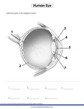
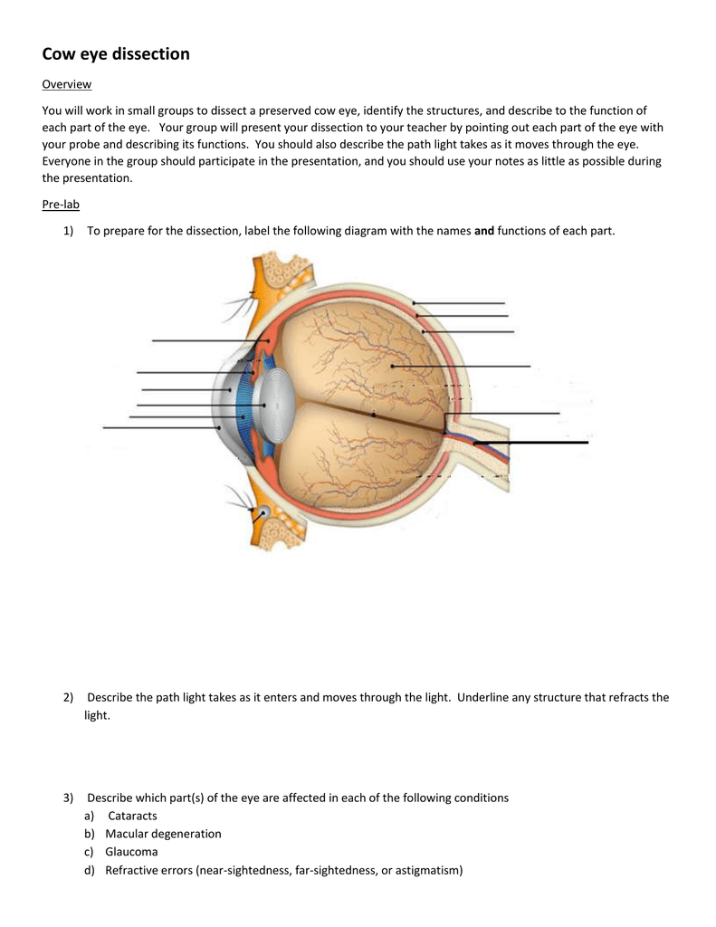
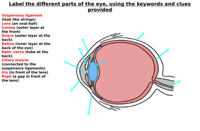
Eye diagram with labels and functions
Diagram of the Eye - Lions Eye Institute Instructions. Click the parts of the eye to see a description for each. Hover the diagram to zoom. Iris. The iris is the coloured part of the eye which surrounds the pupil. It controls light levels inside the eye, similar to the aperture on a camera. The iris contains tiny muscles that widen and narrow the pupil size. Parts of the Eye and Their Functions - Robertson Opt The iris is the area of the eye that contains the pigment which gives the eye its color. This area surrounds the pupil, and uses the dilator pupillae muscles to widen or close the pupil. This allows the eye to take in more or less light depending on how bright it is around you. If it is too bright, the iris will shrink the pupil so that they ... PDF Eye Anatomy Handout - National Eye Institute of light entering the eye. Lens: The lens is a clear part of the eye behind the iris that helps to focus light, or an image, on the retina. Macula: The macula is the small, sensitive area of the retina that gives central vision. It is located in the center of the retina. Optic nerve: The optic nerve is the largest sensory nerve of the eye.
Eye diagram with labels and functions. 2 Way Light Switch Wiring Diagram - House Electrical Wiring Diagram Dec 02, 2015 · A 2 Way Switch Wiring Diagram with Power Feed from the Switch light : ... (833)400-1001.Some of its sophisticated functions are listed here: Setup, Un-installation, as well as Reinstallation of QuickBooks. ... I really needed a way to manage my invoices and to track my financial data. With QuickBooks, I can easily keep a close eye on the ... PDF Parts of the Eye - National Eye Institute | National Eye Institute To understand eye problems, it helps to know the different parts that make up the eye and the functions of these parts. Here are descriptions of some of the main parts of the eye: ... Handout illustrating parts of the eye Keywords: parts of the eye, eye diagram, vitreous gel, iris, cornea, pupil, lens, optic nerve, macula, retina ... Eye Anatomy: 16 Parts of the Eye & Their Functions The following are parts of the human eyes and their functions: 1. Conjunctiva. The conjunctiva is the membrane covering the sclera (white portion of your eye). The conjunctiva also covers the interior of your eyelids. Conjunctivitis, often known as pink eye, occurs when this thin membrane becomes inflamed or swollen. Create a Briliant Process Flow Diagram with Canva Process flow diagrams illustrate how a large complex process is broken down into smaller functions and how these fit together. As visual tools, they can help your team or organization see the bigger picture as well as where they fit into its entirety. Create a process flow any time you want to illustrate the stages of a process.
Eye anatomy: Muscles, arteries, nerves and lacrimal gland - Kenhub Bony cavity within the skull that houses the eye and its associated structures (muscles of the eye, eyelid, periorbital fat, lacrimal apparatus) Bones of the orbit. Maxilla, zygomatic bone, frontal bone, ethmoid bone, lacrimal bone, sphenoid bone and palatine bone. Structure of the eye. Cornea, anterior chamber, lens, vitreous chamber and ... Eye Anatomy: Parts of the Eye and How We See Behind the anterior chamber is the eye's iris (the colored part of the eye) and the dark hole in the middle called the pupil. Muscles in the iris dilate (widen) or constrict (narrow) the pupil to control the amount of light reaching the back of the eye. Directly behind the pupil sits the lens. The lens focuses light toward the back of the eye. Structure and Functions of Human Eye with labelled Diagram The main function is to refract the light along with the lens. Iris: It is the pigmented, coloured portion of the eye, visible externally. The main function of the iris is to control the diameter of the pupil according to the light source. Pupil: It is the small aperture located in the centre of the Iris. Eye anatomy: A closer look at the parts of the eye For more details about specific structures of the eye and how they function, visit these pages: Conjunctiva Of The Eye. Sclera: The White Of The Eye. Cornea Of The Eye. The Uvea Of The Eye. Pupil: Aperture Of The Eye. The Retina: Where Vision Begins. Macula Lutea Of The Eye. Choroid Of The Eye. Lens Of The Eye. Ciliary Body. Eye Muscles ...
› Document › FunctionsListing of NCL Functions by Category Defines eye position, line of sight, up direction, and stereo flag for selected TDPACK routines. tditri: Adds triangles defining an isosurface to a triangle list (for use with selected TDPACK routines). tdlbla: Draws labels for a particular face of a box in 3-space (for use with selected TDPACK routines). tdlblp: Not yet implemented. tdlbls Anatomy of the eye: Quizzes and diagrams - Kenhub Take a look at the diagram of the eyeball above. Here you can see all of the main structures in this area. Spend some time reviewing the name and location of each one, then try to label the eye yourself - without peeking! - using the eye diagram (blank) below. Unlabeled diagram of the eye. Click below to download our free unlabeled diagram of ... Eye pattern - Wikipedia In telecommunication, an eye pattern, also known as an eye diagram, is an oscilloscope display in which a digital signal from a receiver is repetitively sampled and applied to the vertical input, while the data rate is used to trigger the horizontal sweep. It is so called because, for several types of coding, the pattern looks like a series of eyes between a pair of rails. Microscope Types (with labeled diagrams) and Functions A compound microscope: Is used to view samples that are not visible to the naked eye. Uses two types of lenses - Objective and ocular lenses. Has a higher level of magnification - Typically up to 2000x. Is used in hospitals and forensic labs by scientists, biologists and researchers to study micro organisms. Compound microscope labeled diagram.
Labelling the eye — Science Learning Hub Labelling the eye. Use this interactive to label different parts of the human eye. Drag and drop the text labels onto the boxes next to the diagram. Selecting or hovering over a box will highlight each area in the diagram. The human eye has several structures that enable entering light energy to be converted to electrochemical energy.
CUT-AND-ASSEMBLE PAPER EYE MODEL - ellenjmchenry.com THE HUMAN EYE 1) OPTIC NERVE: takes electrical signals to the brain. Notice that the retina’s blood supply comes in through the center of the optic nerve. 2) FOVEA: focal point, the center of your vision 3) MACULA: the area around the fovea 4) RETINA: the back of the inside of the eyeball (This is where the light-sensitive rods and cones are located.)
byjus.com › biology › human-heartHuman Heart - Anatomy, Functions and Facts about Heart Following are the main functions of the heart: One of the primary functions of the human heart is to pump blood throughout the body. Blood delivers oxygen, hormones, glucose and other components to various parts of the body, including the human heart.
Fluorescence - Wikipedia Fluorescence is the emission of light by a substance that has absorbed light or other electromagnetic radiation.It is a form of luminescence.In most cases, the emitted light has a longer wavelength, and therefore a lower photon energy, than the absorbed radiation. A perceptible example of fluorescence occurs when the absorbed radiation is in the ultraviolet …

draw a diagram of the human eye as seen in a vertical section and label the part which suits the ...
Eye Diagram With Labels and detailed description - BYJUS Iris is the coloured part of the eye and controls the amount of light entering the eye by regulating the size of the pupil. The lens is located just behind the iris. Its function is to focus the light on the retina. The optic nerve transmits electrical signals from the retina to the brain. Pupil is the opening at the centre of the iris.

Male Anatomy Diagram Unlabeled : Sc 912 L 16 13 Reproductive System - Blank ear diagram human ...
Eye anatomy and function - AboutKidsHealth A clear lens, located behind the pupil, acts like a camera lens by focusing light onto the retina at the back of the eye. The retina is a light-sensitive inner lining at the back of the eye. Ten different layers of cells work together in the retina to detect light and turn it into electrical impulses. Anatomy of the eye: 3/4 view.
Labelled Diagram of Human Eye, Explanation and Function - VEDANTU The basic functions of Rods and Cones are conscious light perception, color differentiation and depth perception. The human eye is capable of distinguishing between about 10 million colors, and it can also detect a single photo. The human eye is a part of the sensory nervous system. Labeled Diagram of Human Eye

May I have a simplest diagram of an eye, Please - Science - The Human Eye and the Colourful ...
Label Parts of the Human Eye - University of Dayton Label Parts of the Human Eye. Select One Anterior Chamber Ciliary Body Cornea Fibrous Tunic Iris Lateral Rectus Muscle Lens Medial Rectus Muscle Optic Disk Optic Nerve Pupil Retina Vascular Tunic Vitreous Nerve. Select One Anterior Chamber Ciliary Body Cornea Fibrous Tunic Iris Lateral Rectus Muscle Lens Medial Rectus Muscle Optic Disk Optic ...
Human Eye Diagram, How The Eye Work -15 Amazing Facts of Eye The shark has even been used in human eye surgery! FACT 4 The length of our eyes are about 1 inch across and weigh about 0.25 ounce. FACT 5 Our eyeballs stay the same size forever but our nose and ears continue to grow. FACT 6 Eyes are the second most complex organ after the brain.
› help › commGenerate eye diagram - MATLAB eyediagram - MathWorks Description. eyediagram (x,n) generates an eye diagram for signal x, plotting n samples in each trace. The labels on the horizontal axis of the diagram range between -1/2 and 1/2. The function assumes that the first value of the signal and every n th value thereafter, occur at integer times. eyediagram (x,n,period) sets the labels on the ...
The Eye Diagram: What is it and why is it used? The eye diagram is used primarily to look at digital signals for the purpose of recognizing the effects of distortion and finding its source. To demonstrate using a Tektronix MDO3104 oscilloscope, we connect the AFG output on the back panel to an analog input channel on the front panel and press AFG so a sine wave displays.
Labeled Eye Diagram | Eye anatomy diagram, Eye anatomy ... - Pinterest accessory structures of the eye, extrinsic eye muscles, anatomy of the eyeball and microscopic anatomy of the retina. The skeletal system consists of bones and their associated connective tissues, including cartilage, tendons, and ligaments. It consists of dynamic, living tissues that are capable of growth, detect pain stimuli, adapt to stress ...
rsscience.com › stereo-microscopeParts of Stereo Microscope (Dissecting microscope) - Rs' Science If you would like to learn optical components of a compound microscope, please visit Compound Microscope Parts – Labeled Diagram and their Functions, and this article. How to use a stereo (dissecting) microscope. Follow these steps to put your stereo microscopes in work: 1. Set your microscope on a tabletop or other flat sturdy surface where ...
Eye muscles and their functions - All About Vision Recti muscles. The eye has four recti muscles, all of which attach to the front half of the eye (anterior to the equator of the eye). These muscles are: Superior rectus muscle. Medial rectus muscle. Lateral rectus muscle. Inferior rectus muscle. Each of the eye's recti muscles originates from the common tendinous ring (sometimes referred to ...
en.wikipedia.org › wiki › Human_eyeHuman eye - Wikipedia Each eye has seven extraocular muscles located in its orbit. Six of these muscles control the eye movements, the seventh controls the movement of the upper eyelid.The six muscles are four recti muscles – the lateral rectus, the medial rectus, the inferior rectus, and the superior rectus, and two oblique muscles the inferior oblique, and the superior oblique.
Eye Anatomy | Definition, Structure & Functions - iBiologia Diagram of Human Eye with Labelling. Eye Anatomy Complete Physiology of Eye is described below in the given paragraph: The eye is rather like a living Camera. Each eye is a liquid-filled ball 2.5 cm in diameter. At the front of the eye is a clear, round window called the cornea. Behind the cornea is a "lens.
Labelling the eye — Science Learning Hub In this activity, students use online or paper resources to identity and label the main parts of the human eye. By the end of this activity, students should be able to: identify the main parts of the human eye. describe the functions of the different parts of the human eye. Download the Word file (see link below).


Post a Comment for "42 eye diagram with labels and functions"