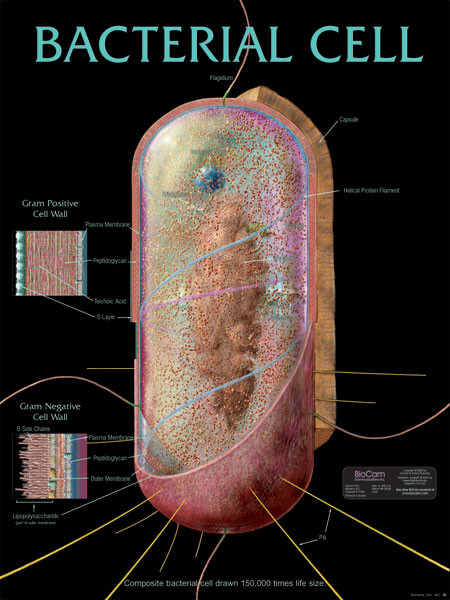
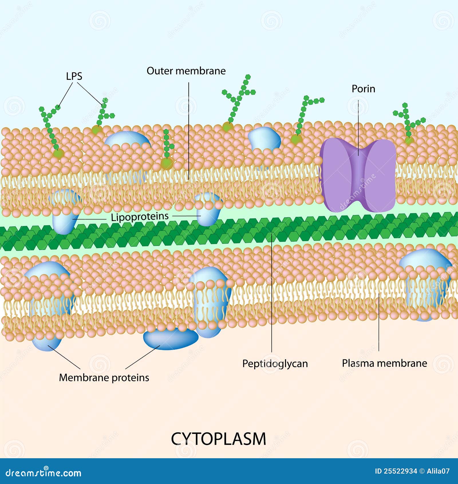
42 bacterial cell picture with labels
Lamisil (terbinafine) vs. Lotrimin (clotrimazole): Antifungal Cream Lamisil. There are no adequate studies in pregnant women. Since nail fungus treatment can be delayed until after pregnancy there is no reason to use oral terbinafine during pregnancy.. Breastfeeding mothers should not use oral terbinafine because terbinafine passes into breast milk.. Lotrimin. Clotrimazole is very poorly absorbed into the blood and the body after … Plant and Animal Cells - Labeled Graphics A compilation of plant and animal cell images with organelles and major structures labeled. Students can print images to help them learn the cell. ... if students missed the lab that day they can view a site with pictures to complete lab handout Plant Cell ... looks at cheek and onion cells. Prokaryote Coloring - color a typical bacteria cell ...
Bacterial cells - Cell structure - Edexcel - GCSE Combined Science ... Bacterial cells Bacteria are all single-celled. The cells are all prokaryotic. This means they do not have a nucleus or any other structures which are surrounded by membranes. Larger bacterial...
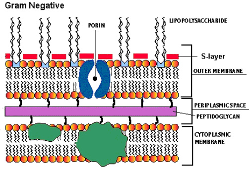
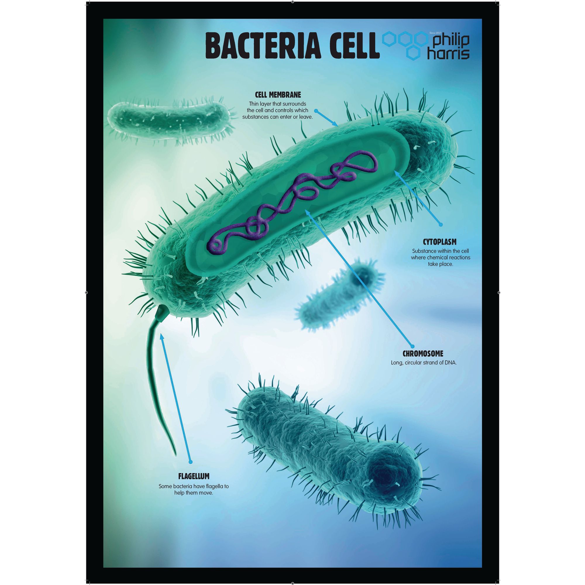
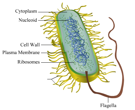
Bacterial cell picture with labels
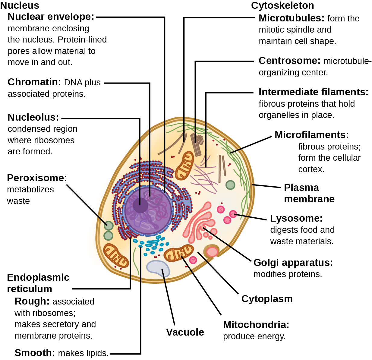
Bacterial Cell Structure Labeling Diagram | Quizlet Bacterial Cell Structure Labeling STUDY Learn Flashcards Write Spell Test PLAY Match Gravity Created by adkelly22 Terms in this set (16) Cytoplasm Water-based solution filling the entire cell Ribosomes Tiny particles composed of protein and RNA that are the sites of protein synthesis Nucleoid Composed of condensed DNA molecules. Cell Membrane Identifying Medical Diagnoses and Treatable Diseases by Image ... - Cell Feb 22, 2018 · Binary comparison of bacterial and viral pneumonia resulted in a test accuracy of 90.7%, with a sensitivity of 88.6% and a specificity of 90.9% (Figures 6C and 6D). The area under the ROC curve for distinguishing bacterial and viral pneumonia was 94.0% (Figure 6F). Animal Cell Labeled Diagram Pictures, Images and Stock Photos Labeled educational bacteria internal structure scheme. Biological blue green algae diagram with carboxysome, thylakoid and phycobilisome parts location inside cell. Animal cell Cells consist of a protoplasm enclosed within a membrane, which contains many biomolecules such as proteins and nucleic acids. Toxoplasma Gondii Structure
Bacterial cell picture with labels. 97,783 Bacteria Cell Stock Photos and Images - 123RF Bacteria Cell Stock Photos And Images 97,783 matches Page of 978 Structure of a bacterial cell. Anatomy of the prokaryote. unicellular organism. Vector diagram for your design, educational, medical, biological and science use Bacteria vector icon isolated on transparent background, Bacteria logo concept Structure of a bacterial cell, labeled. Stock Illustration Download Structure of a bacterial cell, labeled. Stock Illustration and explore similar illustrations at Adobe Stock. Adobe Stock. Photos Illustrations Vectors Videos Audio Templates Free Premium Editorial Fonts. Plugins. 3D. Photos Illustrations Vectors Videos Audio Templates Free Premium Editorial Fonts. Label the Bacterium Cell - EnchantedLearning.com flagellum - A long whip-like structure used for locomotion (movement). Some bacteria have more than one flagellum. pili - (singular is pilus) Hair-like projections that allow bacterial cells to stick to surfaces and transfer DNA to one another. plasma membrane - A permeable membrane located within the cell wall. 2,277 Animal cell labeled Images, Stock Photos & Vectors - Shutterstock 2,277 animal cell labeled stock photos, vectors, and illustrations are available royalty-free. See animal cell labeled stock video clips Image type Orientation Color People Artists More Sort by Biology Animals and Wildlife Healthcare and Medical Cooking Software cell eukaryote in vitro experiment cell culture Turn on AI Powered Search
Erebosis, a new cell death mechanism during homeostatic … 25.04.2022 · It has been believed that gut enterocytes continuously die through apoptosis. However, this study shows that gut enterocytes die through a novel cell death mechanism, named erebosis. Erobotic cells lack the characteristic features of apoptotic, necrotic or autophagic cell death; instead they lose their cytoskeleton, cell adhesion and organelles, and their nuclei … 600+ Free Bacteria & Virus Images - Pixabay Find images of Bacteria. Free for commercial use No attribution required High quality images. Images. Images. Photos. Vector graphics. Illustrations. Videos. Users. Search options ... Find your perfect picture for your project. 639 Free images of Bacteria / 7 ‹ › ... Animal Cell - Free printable to label + Color -kidCourses.com Can you label and color these important parts of the animal cell?. NUCLEUS control center for cell (cell growth, cell metabolism, cell reproduction). NUCLEOLUS synthesizes rRNA. RIBOSOMES the site of protein building, this is where translation takes place (mRNA in language of nucleic acids is translated into the language of amino acids). RER (Rough Endoplasmic Reticulum) synthesizes proteins ... Bacteria in Microbiology - shapes, structure and diagram Bacterial endospores layers Bacteria cells are the smallest living cells that are known; even though viruses are smaller than bacteria, viruses are not living cells. There are different types of bacteria with various sizes, shapes, and structures. The bacteria shapes, structure, and labeled diagrams are discussed below. Sizes
Bacteria Labeled Stock Illustrations - 223 Bacteria Labeled Stock ... Bacteria Labeled Stock Illustrations - 223 Bacteria Labeled Stock Illustrations, Vectors & Clipart - Dreamstime Bacteria Labeled Illustrations & Vectors Most relevant Best selling Latest uploads Within Results People Pricing License Media Properties More Safe Search 223 bacteria labeled illustrations & vectors are available royalty-free. Next page 3 Common Bacteria Shapes - ThoughtCo Bacteria Shapes The three basic shapes of bacteria include cocci (blue), bacilli (green), and spirochetes (red). PASIEKA/Science Photo Library/Getty Images By Regina Bailey Updated on August 20, 2019 Bacteria are single-celled, prokaryotic organisms that come in different shapes. Bacteria Labeled Diagram Stock Vector Image & Art - Alamy Download this stock vector: Bacteria Labeled Diagram - EG0XT7 from Alamy's library of millions of high resolution stock photos, illustrations and vectors. Pooled CRISPR screening with single-cell transcriptome readout 18.01.2017 · CROP-seq enables pooled CRISPR screens for complex transcriptome signatures by making gRNA expression detectable in single-cell RNA sequencing. CRISPR-based genetic screens are accelerating ...
Mouse oocytes develop in cysts with the help of nurse cells: Cell 26.05.2022 · These studies provide a much clearer picture at both the cellular and molecular levels of how oocytes develop in germline cysts. Unactivated nurse cells strongly resemble pro-oocytes and follow a very similar program of gene expression. However, once an individual nurse cell receives a local signal from within its cyst, its gene expression changes quickly, and it …
Cell Size and Scale - University of Utah Smaller cells are easily visible under a light microscope. It's even possible to make out structures within the cell, such as the nucleus, mitochondria and chloroplasts. Light microscopes use a system of lenses to magnify an image. The power of a light microscope is limited by the wavelength of visible light, which is about 500 nm.
Draw a labelled diagram of a bacterial cell. - Careers360 Buy Now NEET Foundation + Knockout NEET 2024 (Easy Installment) Personalized AI Tutor and Adaptive Time Table, Self Study Material, Unlimited Mock Tests and Personalized Analysis Reports, 24x7 Doubt Chat Support,.
50 Striking Microscopic Images of Viruses and Bacteria Click through the slideshow above to see 50 striking electron micrographs of some of the world's most dangerous and deadly disease-causing viruses and bacteria. Know your flu risk. Check out the ...
Manuka Honey: Medicinal Uses, Benefits, and Side Effects Feb 20, 2021 · Health Research Board: A Picture of Health 2008: "Healing with honey." Jull, A. British Journal of Surgery. 2008; vol 95: pp 175-182. Natural Medicines Comprehensive Database.
Animal Cell Labeled Pictures, Images and Stock Photos Labeled educational bacteria internal structure scheme. Biological blue green algae diagram with carboxysome, thylakoid and phycobilisome parts location inside cell. Animal cell Cells consist of a protoplasm enclosed within a membrane, which contains many biomolecules such as proteins and nucleic acids. Toxoplasma Gondii Structure

Image result for illustrations of a cell and different components | Organelles, Eukaryotic cell ...
Powerful single-cell transcriptome analysis in a simple UI Empowering collaboration in annotating cells, discovering unknown cell populations, cell states or cell interactions is crucial to draw the best picture ever of the highly heterogenous tumor microenvironment. Users have access to a growing public as well as their private knowledge base, which makes the BioTuring platform a swiss-army-knife for better understanding cells at the …
Bacteria Label Teaching Resources | Teachers Pay Teachers Plant Cell, Animal Cell, Bacteria Cell Structure Science Poster Labels Anatomy. by. Mrs Wonder's Classroom. $10.00. Zip. Bright colorful set of 3 science printable posters. Plant, Animal and Bacteria cell structure with labels to be used as educational art for any kid's playroom, classroom, Montessori or homeschooling areas.Science Printable ...
Qualitative and Quantitative Analysis in Microbiology For example, recording a moving picture image of the moving cells is used to determine their speed of movement, and whether the presence of a compound acts as an attractant or a repellant to the microbes. Bacterial growth is another area that can yield qualitative or quantitative information. Water analysis for the bacterium Escherichia coli provides an example. A …
PHOTO GALLERY OF BACTERIA - Microbiology in pictures (the cells stain a weak Gram-negative) Microscopic appearance: Spirochetes: Oxygen relationship: microaerobic: Motility: motile: Catalase test:-Oxidase test:- ... Colonies of various bacteria. Bacteria photos. PICTURE OF THE MONTH. BACTERIA 2013 DECEMBER. DISK DIFFUSION METHOD FOR TESTING OF ANTIBIOTIC SUSCEPTIBILITY OF BACTERIA:
Structure of Bacterial Cell (With Diagram) - Biology Discussion It is a tough and rigid structure of peptidoglycan with accessory specific materials (e.g. LPS, teichoic acid etc.) surrounding the bacterium like a shell and lies external to the cytoplasmic membrane. It is 10-25 nm in thickness. It gives shape to the cell. Nucleus: The single circular double-stranded chromosome is the bacterial genome.
Bacteria Cell Structures with Labels Stock Vector - Dreamstime Get 15 images free trial Bacteria Cell Structures with labels Royalty-Free Vector Bacterial cell structures labeled on a bacillus cell with nucleoid DNA and ribosomes. External structures include the capsule, pili, and flagellum. Morphology of internal structures of bacteria. cell anatomy bacteria, prokaryotic cell, cell, internal structures,
Different Size, Shape and Arrangement of Bacterial Cells Size of Bacterial Cell. The average diameter of spherical bacteria is 0.5-2.0 µm. For rod-shaped or filamentous bacteria, length is 1-10 µm and diameter is 0.25-1 .0 µm. E. coli , a bacillus of about average size is 1.1 to 1.5 µm wide by 2.0 to 6.0 µm long. Spirochaetes occasionally reach 500 µm in length and the cyanobacterium.
A Labeled Diagram of the Animal Cell and its Organelles A Labeled Diagram of the Animal Cell and its Organelles. There are two types of cells - Prokaryotic and Eucaryotic. Eukaryotic cells are larger, more complex, and have evolved more recently than prokaryotes. Where, prokaryotes are just bacteria and archaea, eukaryotes are literally everything else. From amoebae to earthworms to mushrooms, grass ...
Plant Cell Labeled Pictures, Images and Stock Photos Labeled bacteria internal structure scheme Cyanobacteria vector illustration. Labeled educational bacteria internal structure scheme. Biological blue green algae diagram with carboxysome, thylakoid and phycobilisome parts location inside cell. plant cell labeled stock illustrations ... Data collection during medical research plant cell labeled ...
Interactive Bacteria Cell Model - CELLS alive Pili, Fimbriae: These hollow, hairlike structures made of protein allow bacteria to attach to other cells. A specialized pilus, the sex pilus, allows the transfer of plasmid DNA from one bacterial cell to another. Pili (sing., pilus) are also called fimbriae (sing., fimbria). Flagella: The purpose of flagella (sing., flagellum) is motility.
Levaquin (levofloxacin) Antibiotic Side Effects, Uses & Dosage Levofloxacin (Levaquin - a discontinued brand) is a prescription drug used to treat bacterial infections of the sinuses, skin, lungs, ears, airways, bones, and joints. Side effects include nausea, vomiting, diarrhea, headache, and constipation. Read the warnings, drug interactions, dosage, and pregnancy and breastfeeding safety information.





Post a Comment for "42 bacterial cell picture with labels"